The cause of many common foot problems, such as plantar fasciitis, tendonitis & metatarsalgia is the combination of tight muscles and overpronation
Full Muscular System Description [Continued from above] . . . Muscular System Anatomy. Muscle Types There are three types of muscle tissue: Visceral, cardiac, and

The Skeletal System – Extensive anatomy images and detailed descriptions allow you to learn all about the bones of the human skeleton, as well as ligaments.
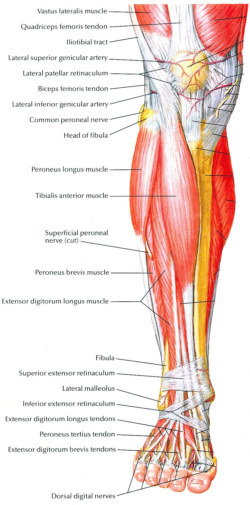
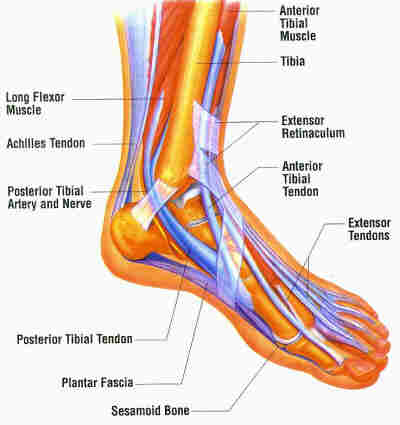
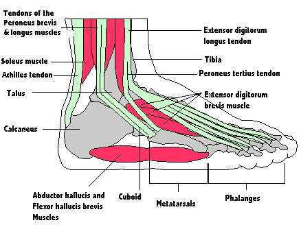
A network of more than 100 muscles, tendons, and ligaments helps give the foot its strength, mobility, and versatility.


The midfoot is the middle region of the foot, where a cluster of small bones forms an arch on the top of the foot. From this cluster, five long bones (metatarsals
Muscle: Origin: Insertion: Function: Location: For images of the muscle, click on each link under location. Abductors (tensor fasciae latae, gluteus medius, gluteus
The action potential When chemicals contact the surface of a neuron, they change the balance of ions (electrically charged atoms) between the inside and outside of
The following diagram illustrates the actions of the terms adduction, abduction, flexion and extension at the different joints. This is important to understand the


WebMD provies information about the antomy of the muscle including the function, conditions affecting the including injuries, and much more.


The blood vessels of the foot supply oxygenated blood to muscles and return blood back to the heart. The nerves send sensory information to the brain.
